
Intraoral Camera in Layton, UT
Intraoral camera imaging helps patients see what our dentist and team see. This page explains what an intraoral camera in Layton, Utah, is, how it works, the benefits of this dental imaging technology, and what to expect during an appointment.
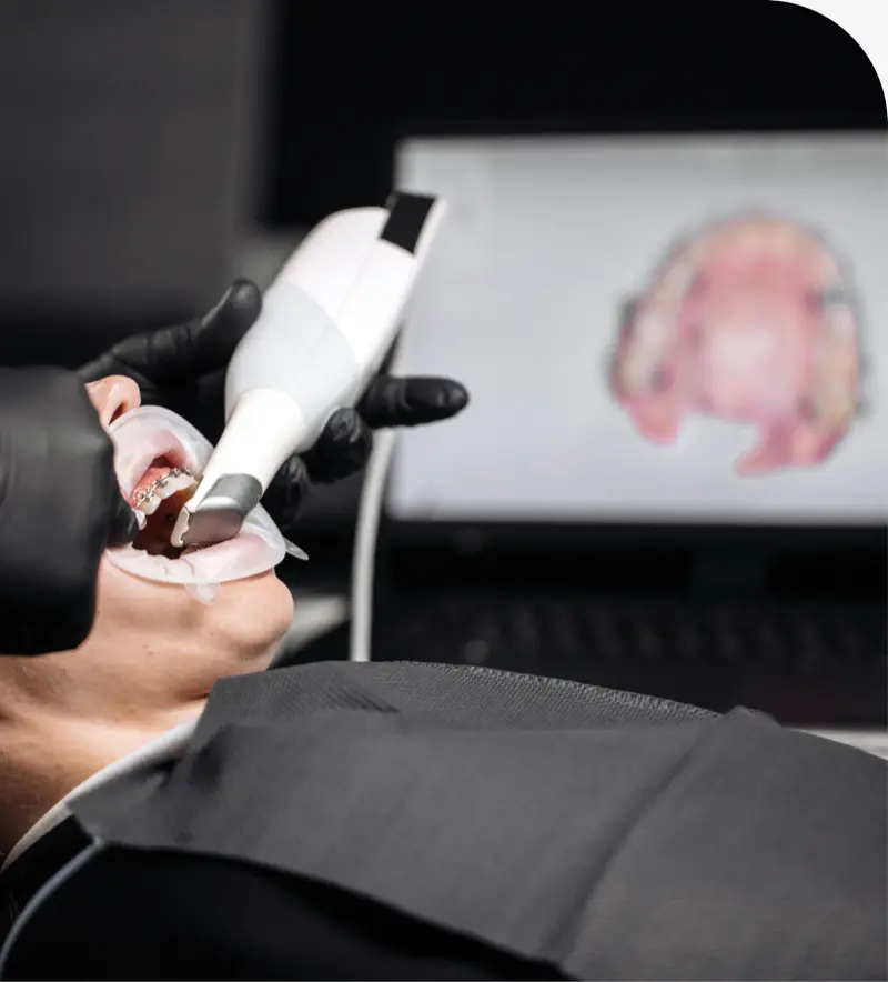
Intraoral Camera Explained
An intraoral camera is a small, handheld device with a bright light and a high-resolution lens. It captures close-up color photos and short videos of your teeth and gums. Images appear on a chairside screen in real time, so concerns such as cavities, cracks, worn fillings, plaque buildup, and early gum inflammation are easier to spot and discuss.
This tool does not emit radiation and does not replace necessary X-rays. Instead, it complements X-rays by showing surface details that X-rays may not reveal. Disposable, single-use covers protect your health and keep the process clean from start to finish.
Benefits of Intraoral Camera Imaging
- Clearer diagnosis through magnified, color images of teeth and gums.
- Early detection of cavities, fractures, and wear before they worsen.
- Patient education with on-screen visuals that make findings easy to understand.
- Minimally invasive imaging without discomfort, heat, or radiation.
- Accurate documentation for monitoring changes over time.
- More precise treatment planning for fillings, crowns, and gum care.
- Helpful visuals for insurance documentation when needed.
How Intraoral Camera Imaging Works
During a routine exam, our dentist or clinical team will gently position the camera tip near each tooth surface. The light illuminates hard-to-see areas, and the lens magnifies fine details. Images display instantly on a chairside monitor for review.
Typical steps include:
- Initial scan to survey teeth and gums and identify areas that need a closer look.
- Targeted images of specific spots showing cracks, decay, or gum changes.
- Review and explanation of what the images show, plus options for care.
- Saving images to your record to track changes and outcomes over time.
What to Expect During Your Appointment
Most image capture takes only a few minutes and feels similar to a small mirror touching the teeth. There is no special preparation required, though removing heavy lipstick can keep images clear.
Expect our dentist to use the photos to explain findings, discuss prevention, and outline treatment choices if needed. The camera is especially useful for monitoring gum health, evaluating chips or fractures, confirming the fit of new fillings and crowns, and documenting before-and-after results.
Did You Know?
Intraoral cameras magnify your teeth up to 25x, letting you see exactly what your dentist sees during your exam.
Got Questions?
Frequently Asked Questions
Yes. It uses visible light only and produces no radiation. A single-use barrier covers the camera for infection control.
No. X-rays show internal structures and areas between teeth, while intraoral photos show surface details. Together, they give a more complete picture.
It helps reveal cavities, cracks, worn enamel, failing fillings, plaque and tartar buildup, and signs of gum irritation.
Yes. Photos are stored in your digital chart so changes can be compared over time and referenced during future visits.
No. Image capture is quick and often shortens explanations because you can see the condition immediately on the screen.
Yes. The visuals help children and parents understand hygiene tips, early cavities, and orthodontic considerations in a simple, engaging way.
No. The camera is smooth and small. It touches the teeth lightly and should not cause discomfort.
Want to See Your Smile Up Close?
Call (801) 444-9090 or contact 4 Dental Health to schedule an exam with intraoral camera imaging in Layton, UT with Dr. Steven Christensen.